Veterinary Diagnostics
The Georgia Electron Microscopy Facility provides diagnostic services to the veterinary and medical professions. We have the unique opportunity to work closely with the Pathology Department at the College of Veterinary Medicine. If extra pathological support is needed for a particular case, we do enlist one of our pathologists who would have the best expertise in a particular area of tissue ultrastructure.
Before sending or submitting specimens, please make contact by emailing the Georgia Electron Microscopy Facility. Turnaround times will vary depending on the amount of veterinary work being done at any one time within the facility.
Our new address is ISTEM Research Bldg 1, Suite 1000, 302 East Campus Road, Athens, GA 30602. For more information, please contact the EM Lab Coordinator for Veterinary Services, Mary Ard .

Services Offered
*Diagnostic Services *Research Services *Instruction and Training
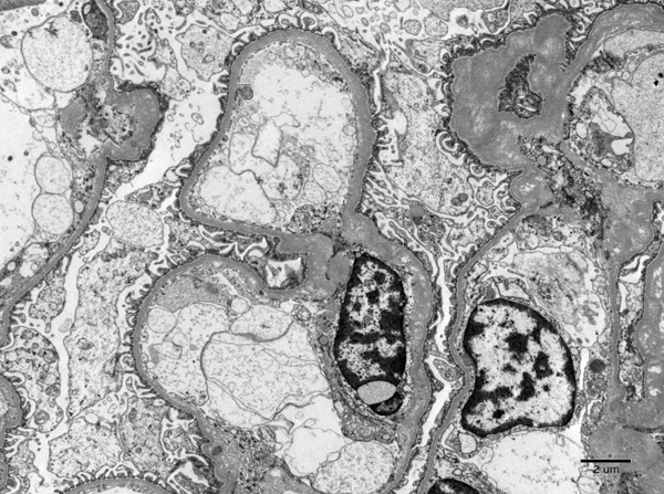
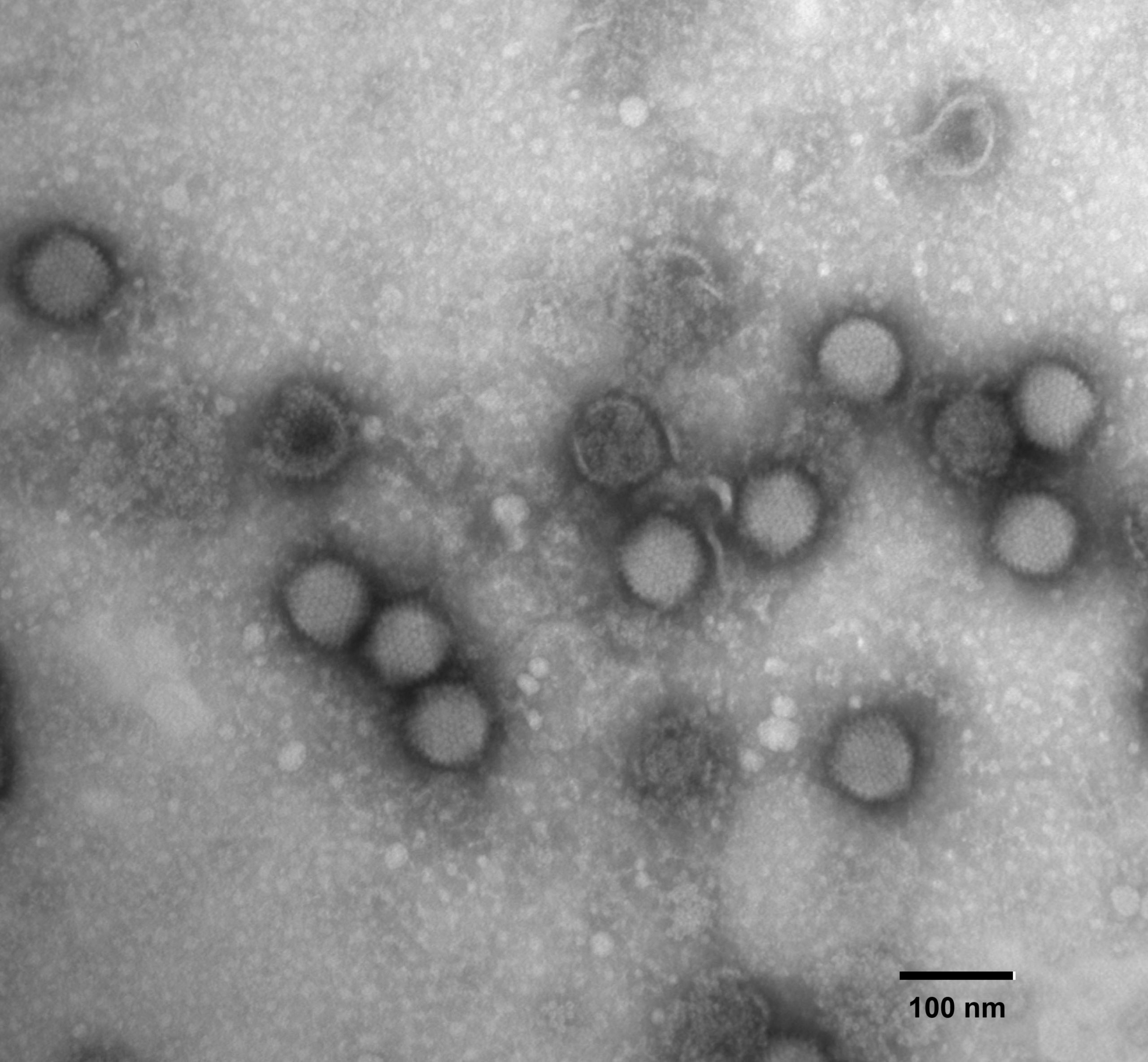
A complete range of conventional preparative services are offered, allowing investigators to submit fresh or fixed tissues and receive end results as requested (i.e. pathologist evaluation and/or digital imaging) within a reasonable turnaround time. Services include Transmission Electron Microscopy (TEM), Negative Staining/Negative Contrast, and Scanning Electron Microscopy (SEM). See our fee schedule for available services and pricing.
Diagnostic Services
Diagnostic services are available through the Department of Pathology and the Veterinary Diagnostic Laboratories, in which electron microscopy is essential for correct diagnosis. Their services will include a pathologist who will take on the diagnostic case. Outside veterinary clinics, other diagnostic facilities, and universities requiring electron microscopy for Veterinary Diagnostics are welcome to contact GEM to inquire about how we can provide services for you. Contact us by email: gem@uga.edu or maryard@uga.edu
Primary Ciliary Dyskinesia information
Research Services
Research in electron microscopy is done for various departments within the College of Veterinary Medicine and other units throughout the UGA campus. EM research is available to surrounding research facilities as well. More information can be found at the Georgia Electron Microscopy Facility’s homepage.
Instruction and Training
On-site instruction and training is available to most individuals who would like to utilize the facility for their own research and casework. All instruction and training is done at the Georgia Electron Microscopy Facility.
Workshops on biological specimen preparation for TEM are also available. More information is located here.
Submission Process
Negative staining for viral confirmation/identification
Tissue processing for Transmission Electron Microscopy (TEM)
Tissue processing for Scanning Electron Microscopy (SEM)
EM Services Submission Form for Diagnostics
Forms must be filled out completely and accompany all samples submitted to the Georgia Electron Microscopy Facility. Priority Software – FBS account registration will be required.
Sample Submission Guidelines
Negative staining for viral confirmation/ identification
Samples such as veterinary cell culture, fecal material, allantoic fluid, fractionated samples (biosafety level 2 or lower) should be submitted fresh in a closed container or syringe with needle detached or protected. Samples of biosafety level 3 should be inactivated (we prefer paraformaldehyde over UV for inactivation methods) before sending to the GEM Lab. Clinic # or other identification must be written on the container/syringe to match what is on the submission form, or the sample will not be accepted. Tissue samples for negative staining should be submitted fresh, double contained, and the GEM Lab should be notified prior to submission. There are extra charges for concentrating fecal material and homogenizing tissue samples. Preparation and interpretation will take 8–72 hours depending on workload.
Tissue processing for Transmission Electron Microscopy (TEM)
Samples should be submitted fixed in an approved EM fixative*. Samples should be submitted in pieces no larger than 2mm at any one length. Fixed tissue should be kept refrigerated once collected. Clinic # or other identification must be written on the container as well as the type of fixative used to fix the sample. Processing the fixed tissue into plastic blocks will take 5–7 working days.
* An approved EM fixative contains a 1.5%–3% glutaraldehyde solution in a stable buffer pH 6.8–7.3. It may also contain formaldehyde of up to 4%. Appropriate fixatives are available in the GEM Lab.
Tissue processing for Scanning Electron Microscopy (SEM)
Samples should be submitted fixed in an approved EM fixative*. Sample can be up to 1cm (1.5 in) at any one length, but be aware that the ratio of fixative: sample should be 10:1 (v/v). Fixed tissue should be kept refrigerated once collected. Clinic # or other identification must be written on the container as well as the type of fixative used to fix the sample. Processing the fixed tissue for SEM viewing will normally take 3–5 working days.
* An approved EM fixative contains a 1.5%–3% glutaraldehyde solution in a stable buffer pH 6.8–7.3. It may also contain formaldehyde of up to 4%. Appropriate fixatives are available in the GEM Lab.

